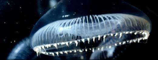
Green Fluorescent Protein (GFP) has existed for more than one hundred and sixty million years in one species of jellyfish, Aequorea victoria. The protein is found in the photoorgans of Aequorea, see picture below right. GFP is not responsible for the glow often seen in pictures of jellyfish – that “fluorescence” is actually due to the reflection of the flash used to photograph the jellies.
Aequorea victoria
Aequorea victoria photoorgans
(Photocredit: Steve Haddock and his bioluminescence web page, Monterey Bay Aquarium Research Institute)
In Aequorea victoria a protein called aequorin releases blue light upon binding with calcium. This blue light is then totally absorbed by the GFP, which in turn gives off the green light.
In 1994 GFP was cloned. Now GFP is found in laboratories all over the world where it is used in every conceivable plant and animal. Flatworms, algae, E. coli and pigs have all been made to fluoresce with GFP.
The importance of GFP was recognized in 2008 when the Nobel Committee awarded Osamu Shimomura, Marty Chalfie and Roger Tsien the Chemistry Nobel Prize “for the discovery and development of the green fluorescent protein, GFP.”
Why is it so popular? Well, I like to think of GFP as the microscope of the twenty-first century. Using GFP we can see when proteins are made, and where they can go. This is done by joining the GFP gene to the gene of the protein of interest so that when the protein is made it will have GFP hanging off it. Since GFP fluoresces, one can shine light at the cell and wait for the distinctive green fluorescence associated with GFP to appear.
Check out some of the visually arresting photographs, shown below, that have been taken of fluorescently labeled proteins. Although the pictures are amazing, what they tell us is even more fantastic. Check out Cool Uses for more info.
On this site we will present you with an introduction to GFP. If you are interested in reading more about GFP, there is always Glowing Genes and Illuminating Disease.